
Другие части тела:
Кроссворд «Части тела – Внутренние органы»
Скачать карточки «Внутренние органы»
Parasympathetic innervation to the ascending colon is supplied by the vagus nerve. Sympathetic innervation is supplied by the splanchnic nerves that join the celiac ganglia. Most of the digestive tract is innervated by the two large celiac ganglia, with the upper part of each ganglion joined by the greater splanchnic nerve and the lower parts joined by the lesser splanchnic nerve. It is from these ganglia that many of the gastric plexuses arise.
Dietary life rules, Japan, Edo period Illustrating the ill effects of drinking alcohol on the digestive system.
Historical depiction of the digestive system, 17th century Persia
In the 19th century it was accepted that chemical processes were involved in the process of digestion. Physiological research into secretion and the gastrointestinal tract was pursued with experiments undertaken by Claude Bernard, Rudolph Heidenhain and Ivan Pavlov.
Arteries and veins around the pancreas and spleen
The digestive system is supplied by the celiac artery. The celiac artery is the first major branch from the abdominal aorta, and is the only major artery that nourishes the digestive organs.
There are three main divisions – the left gastric artery, the common hepatic artery and the splenic artery.
The celiac artery supplies the liver, stomach, spleen and the upper 1/3 of the duodenum (to the sphincter of Oddi) and the pancreas with oxygenated blood. Most of the blood is returned to the liver via the portal venous system for further processing and detoxification before returning to the systemic circulation via the hepatic veins.
The next branch from the abdominal aorta is the superior mesenteric artery, which supplies the regions of the digestive tract derived from the midgut, which includes the distal 2/3 of the duodenum, jejunum, ileum, cecum, appendix, ascending colon, and the proximal 2/3 of the transverse colon.
The final branch which is important for the digestive system is the inferior mesenteric artery, which supplies the regions of the digestive tract derived from the hindgut, which includes the distal 1/3 of the transverse colon, descending colon, sigmoid colon, rectum, and the anus above the pectinate line.
Each part of the digestive system is subject to a wide range of disorders many of which can be congenital. Mouth diseases can also be caused by pathogenic bacteria, viruses, fungi and as a side effect of some medications. Mouth diseases include tongue diseases and salivary gland diseases. A common gum disease in the mouth is gingivitis which is caused by bacteria in plaque. The most common viral infection of the mouth is gingivostomatitis caused by herpes simplex. A common fungal infection is candidiasis commonly known as thrush which affects the mucous membranes of the mouth.
Stomach diseases are often chronic conditions and include gastroparesis, gastritis, and peptic ulcers.
A number of problems including malnutrition and anemia can arise from malabsorption, the abnormal absorption of nutrients in the GI tract. Malabsorption can have many causes ranging from infection, to enzyme deficiencies such as exocrine pancreatic insufficiency. It can also arise as a result of other gastrointestinal diseases such as coeliac disease. Coeliac disease is an autoimmune disorder of the small intestine. This can cause vitamin deficiencies due to the improper absorption of nutrients in the small intestine. The small intestine can also be obstructed by a volvulus, a loop of intestine that becomes twisted enclosing its attached mesentery. This can cause mesenteric ischemia if severe enough.
«Digestive system» and «alimentary system» redirect here. For digestive systems of non-human animals, see Digestion.
The human digestive system consists of the gastrointestinal tract plus the accessory organs of digestion (the tongue, salivary glands, pancreas, liver, and gallbladder). Digestion involves the breakdown of food into smaller and smaller components, until they can be absorbed and assimilated into the body. The process of digestion has three stages: the cephalic phase, the gastric phase, and the intestinal phase.
The first stage, the cephalic phase of digestion, begins with secretions from gastric glands in response to the sight and smell of food. This stage includes the mechanical breakdown of food by chewing, and the chemical breakdown by digestive enzymes, that takes place in the mouth. Saliva contains the digestive enzymes amylase, and lingual lipase, secreted by the salivary and serous glands on the tongue. Chewing, in which the food is mixed with saliva, begins the mechanical process of digestion. This produces a bolus which is swallowed down the esophagus to enter the stomach.
The second stage of digestion begins in the stomach with the gastric phase. Here the food is further broken down by mixing with gastric acid until it passes into the duodenum, the first part of the small intestine.
The third stage begins in the duodenum with the intestinal phase, where partially digested food is mixed with a number of enzymes produced by the pancreas. Digestion is helped by the chewing of food carried out by the muscles of mastication, the tongue, and the teeth, and also by the contractions of peristalsis, and segmentation. Gastric acid, and the production of mucus in the stomach, are essential for the continuation of digestion.
Peristalsis is the rhythmic contraction of muscles that begins in the esophagus and continues along the wall of the stomach and the rest of the gastrointestinal tract. This initially results in the production of chyme which when fully broken down in the small intestine is absorbed as chyle into the lymphatic system. Most of the digestion of food takes place in the small intestine. Water and some minerals are reabsorbed back into the blood in the colon of the large intestine. The waste products of digestion (feces) are defecated from the rectum via the anus.
The digestive system, or gastrointestinal tract, begins with the mouth, where food enters the body, and ends with the anus, where solid waste material leaves the body. The primary functions of the organs of the digestive system are threefold.
First, complex food material which is taken into the mouth (ingestion) must be digested, or broken down, mechanically and chemically, as it travels through the gastrointestinal tract. Complex proteins are digested to simpler amino acids; complicated sugars are reduced to simple sugars, such as glucose; and large fat molecules are broken down to fatty acids and triglycerides.
Second, the digested food must be absorbed by passage through the walls of the small intestine into the bloodstream so that the valuable energy-carrying nutrients (sugars, amino acids, fatty acids) can travel to all the cells of the body. Within the cells, sugars and fatty acids can be burned in the presence of oxygen (catabolism), thereby releasing the energy stored in the food matter. Amino acids are used by the cells to build large protein molecules (anabolism) necessary for growth and development.
The third function of the gastrointestinal tract is to eliminate the solid waste materials which are unable to be absorbed by the small intestine. The solid wastes (feces) are concentrated in the large intestine and finally passed out of the body through the anus.
a) 1. deglutition; 2. absorption; 3. mastication; 4. digestion; 5. peristalsis; 6. excretion; 7. anastomosis; 8. regurgitation.
b) 1. bringing food back up the gastrointestinal tract; 2. breakdown of complex substances; 3. new opening up between two hollow organs or fibers; 4. contraction and relaxation of muscles to propel food along the gastrointestinal tract; 5. formation of wastes and removal from the body; 6. passage of simple nutrients into the bloodstream; 7. swallowing; 8. chewing.
Exercise 2.Translate into English:
Шлунково-кишковий тракт; клітини; жирні кислоти; ковтання; амінокислоти; кисень; усувати; тонка кишка; товста кишка; живильні речовини.
The wall of the greater part of the digestive tract consists of three coats: internal — mucous, middle — muscular, and the external — serous. The mucous coat is lined with the epithelium outside which is a connective tissue with a thin layer of smooth muscle fibres. The mucous coat is pink in colour because it has many blood vessels. The numerous small glands in this coat secrete a viscous coat of the digestive tract. It facilitates the movement of food and protects the mucous coat from the damage by solid particles of food and various chemical substances. One must remember that the mucous coat of the digestive tract begins with the esophagus, contains lymph nodules which also have a protective function.
The greater part of the muscular coat of the digestive tract consists of two layers: an internal layer with circular muscle fibres and an external layer with longitudinal muscle fibres. The wall of the pharynx and the superior part of the esophagus, and the tongue and the soft palate all contain striated muscle tissue. It is the muscular coat of the other parts of the digestive tract that consists of smooth muscle tissue. Contractions of the muscular coat move food along the digestive tract.
The serous coat that covers the digestive organs in the abdominal cavity is called the peritoneum. The peritoneum has two layers, visceral and parietal. In the esophagus the serous layer is lacking and the outer coat is fibrous in nature.
The digestive glands secrete digestive juices that contain enzymes and some other substances which take part in the chemical processes of digestion.
In addition to the small glands in the mucous coat of the digestive tract, there are also large glands: the salivary glands, the liver and the pancreas. Though these glands are situated outside the digestive tract, they communicate with it through ducts.
Any part of the digestive tract and the digestive glands are equipped with nerve fibres and their endings. The nerves of the digestive glands regulate the secretion of digestive juices. It is known that the nervous system not only regulates the activity of each organ, but also coordinates their activities.
Стенки большей частью пищеварительного тракта состоит из трех слоев: внутреннего — слизистой оболочки, в середине — мощный, и внешней — обесточиванию потребителей. Слизистые оболочки тонкий слой не будет выровнен в этом случае за пределы которое является соединительной ткани с помощью тонкого слоя гладких мышечных волокон. Слизистые оболочки слой розового цвета из-за многих кровеносных сосудов.В многочисленных небольших кабельных сальников в этот слой нотариальное вязкой покрыть пищеварительного тракта. Она облегчает передвижение и защищает слизистые оболочки слой от повреждения, твердые частицы пищи и различных химических веществ. Надо помнить, что слизистые оболочки слой на желудочно-кишечный тракт начинается с пищевод, содержит лимфатических конкреций, также имеющих защитную функцию.
По большей части мышечной слой на пищеварительный тракт состоит из двух слоев: внутреннего слоя с круглой мышечных волокон и внешнего слоя с продольных мышечных волокон. В стенке зева и превосходная часть пищевода, и на родном языке, и мягкий вкус все содержат вестибулярных рецепторов тканей.Это мощный слой из других частей пищеварительного тракта, которая состоит из гладкой мышечной ткани. Спад в мышечной слой пищу в желудочно.ветровому обесточиванию потребителей в пальто, которое охватывает органов пищеварения в брюшной полости, называется Брюшина. В Брюшина имеет два слоя, висцерального и нижней.В пищевод к летальному исходу слой является недостаточно, и наружный слой, волокнистые характер.ветровому пищеварительные железы нотариальное пищеварительные соки, которые содержат ферменты и некоторых других веществ, которые принимают участие в химических процессов ферментации.ветровому в дополнение к малым железами в слизистой оболочки покрыть пищеварительного тракта, а также большие железы: слюнных железы, печени и поджелудочной железы.Несмотря на то, что эти железы расположены за пределами пищеварительного тракта, они связываются с его через каналы.ветровому какой-либо части пищеварительного тракта и пищеварительные железы есть нервные волокна и их преждевременного завершения заседаний. Нажим на пищеварительные железы регулируют гастриты пищеварительной соки. Известно, что нервная система не только регулирует деятельность каждого органа,Но и координирует их деятельность.
У англичан есть забавное детское стихотворение о том, из чего сделаны мальчики и девочки (What Are Little Boys and Girls Made Of?). Я особенно люблю строчку, в которой говорится, что девочки сделаны из конфет и пирожных, а также всевозможных сластей. Давайте заглянем внутрь нашего тела и узнаем, как системы организма и внутренние органы называются на английском.
Предлагаю отработать правильное произношение слов с помощью следующего видео.
Глаголы, которые описывают процессы, происходящие в нашем организме
Человеческий организм можно сравнить со сложным механизмом, в котором взаимосвязаны все шестеренки и детали. Если что-то выходит из строя, это отражается на функционировании всей системы. Сбои в нашем организме ведут к болезням и осложнениям, чтобы этого избежать, необходимо проходить диагностику. О ее видах вы узнаете из следующей таблицы.
Купить в аптеке нужное лекарство и посоветоваться с аптекарем будет намного легче, если вы внимательно прочитаете статью из нашего блога.
А чтобы не запутаться в анатомических терминах и медицинских понятиях, предлагаю посмотреть видео от преподавателя-носителя. Обратите внимание на полезную лексику и правильное произношение.
Список полезной лексики из видео
Яркий мультфильм на английском поможет вам понять, как устроен организм человека.
ГБОУ СПО «Тольяттинский медколледж»
Методическая разработка практического занятия
Органы пищеварительной системы. The Past Perfect Tense
исциплина «Иностранный язык» (английский)
Специальность: 060604 Лабораторная диагностика (базовая подготовка)
Преподаватель: Семенова Н. В.
занятия для преподавателя
Тема. Органы пищеварительной системы. The Past Perfect Tense.
После изучения данной темы студент должен уметь:
– вести беседу по теме;
– понимать лексические единицы и выражения в потоке речи;
– читать и переводить тексты по изученной теме.
После изучения данной темы студент должен знать:
–органы пищеварительной системы и их функции;
– употребление The Past Perfect Tense;
– лексику по теме.
Воспитательные цели. Изучение данной темы:
– стимулирует интерес студентов к обучению в колледже;
– стимулирует самостоятельность студенческого творчества.
– способствует созданию мотивации на продолжение самостоятельного углубленного изучения дисциплины.
В результате освоения данной темы у студента должны формироваться следующие общие компетенции
Общее время занятия – 2 часа.
занятия: учебный кабинет колледжа «Иностранный язык».
Оснащение занятия: компьютер, мультимедийный проектор, видеофильм, презентация.
План проведения занятия
1. Elaine N. Marieb. Human Anatomy & Physiology. Sixth Edition
2. Kent M. Van De Graaff, Stuart Ira Fox. Concepts of Human Anatomy and Physiology. Third Edition. Wm. C. Brown Publishers.
3. Raymond Murphy. English Grammar in Use. Third Edition. Cambridge University Press.
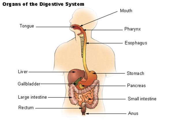
Organs of Digestive System
Food begins its journey through the digestive system in the mouth, also known as the . Inside the mouth are many accessory organs that aid in the digestion of food—the tongue, teeth, and salivary glands. Teeth chop food into small pieces, which are moistened by saliva before the tongue and other muscles push the food into the pharynx.
are 32 small, hard organs found along the anterior and lateral edges of the mouth. Each tooth is made of a bone-like substance called dentin and covered in a layer of enamel—the hardest substance in the body. Teeth are living organs and contain blood vessels and nerves under the dentin in a soft region known as the pulp. The teeth are designed for cutting and grinding food into smaller pieces.
is located on the inferior portion of the mouth just posterior and medial to the teeth. It is a small organ made up of several pairs of covered in a thin, bumpy, skin-like layer. The outside of the tongue contains many rough papillae for gripping food as it is moved by the tongue’s muscles. The taste buds on the surface of the tongue detect taste molecules in food and connect to nerves in the tongue to send taste information to the brain. The tongue also helps to push food toward the posterior part of the mouth for swallowing.
Surrounding the mouth are 3 sets of salivary glands. The salivary glands are accessory organs that produce a watery secretion known as saliva. Saliva helps to moisten food and begins the digestion of carbohydrates. The body also uses saliva to lubricate food as it passes through the mouth, pharynx, and esophagus.
The pharynx, or throat, is a funnel-shaped tube connected to the posterior end of the mouth. The pharynx is responsible for the passing of masses of chewed food from the mouth to the esophagus. The pharynx also plays an important role in the respiratory system, as air from the nasal cavity passes through the pharynx on its way to the larynx and eventually the . Because the pharynx serves two different functions, it contains a flap of tissue known as the that acts as a switch to route food to the esophagus and air to the
is a muscular tube connecting the pharynx to the stomach that is part of the upper gastrointestinal tract It carries swallowed masses of chewed food along its length. At the inferior end of the esophagus is a muscular ring called the loweresophageal sphincter or cardiac sphincter. The function of this sphincter is to close of the end of the esophagus and trap food in the stomach.
is a muscular sac that is located on the left side of the abdominal cavity, just inferior to the . In an average person, the stomach is about the size of their two fists placed next to each other. This major organ acts as a storage tank for food so that the body has time to digest large meals properly. The stomach also contains hydrochloric acid and digestive enzymes that continue the digestion of food that began in the mouth.
is a long, thin tube about 1 inch in diameter and about 10 feet long that is part of the lower gastrointestinal tract. It is located just inferior to the stomach and takes up most of the space in the abdominal cavity. The entire small intestine is coiled like a hose and the inside surface is full of many ridges and folds. These folds are used to maximize the digestion of food and absorption of nutrients. By the time food leaves the small intestine, around 90% of all nutrients have been extracted from the food that entered it.
Liver and Gallbladder. is a roughly triangular accessory organ of the digestive system located to the right of the stomach, just inferior to the diaphragm and superior to the small intestine. The liver weighs about 3 pounds and is the second largest organ in the body. The liver has many different functions in the body, but the main function of the liver in digestion is the production of bile and its secretion into the small intestine. The is a small, pear-shaped organ located just posterior to the liver. The gallbladder is used to store and recycle excess bile from the small intestine so that it can be reused for the digestion of subsequent meals.
is a large gland located just inferior and posterior to the stomach. It is about 6 inches long and shaped like short, lumpy snake with its “head” connected to the duodenum and its “tail” pointing to the left wall of the abdominal cavity. The pancreas secretes digestive enzymes into the small intestine to complete the chemical digestion of foods.
The large intestine is long, thick tube about 2 ½ inches in diameter and about 5 feet long. It is located just inferior to the stomach and wraps around the superior and lateral border of the small intestine. The large intestine absorbs water and contains many symbiotic bacteria that aid in the breaking down of wastes to extract some small amounts of nutrients. Feces in the large intestine exit the body through the anal canal.
Answer the questions
1. Where does the process of digestion begin?
2. What is the hardest tissue in your body?
3. Why do we need the taste buds?
4. What are the functions of saliva?
5. Why do we need the epiglottis?
6. What is the function of cardiac sphincter?
7. What chemicals digest food in the stomach?
8. Where are nutrients absorbed?
9. What are the functions of liver and gallbladder?
10. What is the function of pancreas?
11. Where does the absorption of water occur?
Тест на знание времени Past Perfect
Выберите правильный вариант
1. I (wake up) early and got out of bed.
had woken up
2. I got out of bed an hour later I (wake up).
3. We were late. The meeting (start) an hour before.
4. She was the most delightful person I (ever / meet).
had ever met
5. That morning she (dress), (phone) somebody, and went out.
had dressed, had phoned
6. That morning she went out after she (phone) somebody.
7. He was tired because he (work) hard in the garden all day.
8. The sun (set), it (get) dark, and we went home.
had set, got
9. The Hills were in a hurry, but they (take) a taxi and managed to arrive exactly on time.
B had taken
10. The Hills managed to arrive exactly on time because they (take) a taxi.
I lost the key that he (give) to me
12. We asked Peter to come with us, but he refused. He (already / promise) to play football with his friends.
had already promised
13. I saw a nice kitten when I (open) the basket.
14. After I (write) all my letters, I went to the kitchen to make coffee.
15. I went downstairs because I (hear) the noise.
Ответы к тесту по грамматике
1.a 2b 3.b 4.b 5.a 6.b 7.b 8.a 9.a 10.b 11.a 12.b 13.a 14.b 15.b
There are several organs and other components involved in the digestion of food. The organs known as the accessory digestive organs are the liver, gall bladder and pancreas. Other components include the mouth, salivary glands, tongue, teeth and epiglottis.
A major digestive organ is the stomach. Within its mucosa are millions of embedded gastric glands. Their secretions are vital to the functioning of the organ.
Most of the digestion of food takes place in the small intestine which is the longest part of the GI tract.
There are many specialised cells of the GI tract. These include the various cells of the gastric glands, taste cells, pancreatic duct cells, enterocytes and microfold cells.
3D Medical illustration explaining the oral digestive system
Food enters the mouth where the first stage in the digestive process takes place, with the action of the tongue and the secretion of saliva. The tongue is a fleshy and muscular sensory organ, and the first sensory information is received via the taste buds in the papillae on its surface. If the taste is agreeable, the tongue will go into action, manipulating the food in the mouth which stimulates the secretion of saliva from the salivary glands. The liquid quality of the saliva will help in the softening of the food and its enzyme content will start to break down the food whilst it is still in the mouth. The first part of the food to be broken down is the starch of carbohydrates (by the enzyme amylase in the saliva).
Esophagus shown in yellow passing behind the trachea and the heart
The esophagus, commonly known as the foodpipe or gullet, consists of a muscular tube through which food passes from the pharynx to the stomach. The esophagus is continuous with the laryngopharynx. It passes through the posterior mediastinum in the thorax and enters the stomach through a hole in the thoracic diaphragm—the esophageal hiatus, at the level of the tenth thoracic vertebra (T10). Its length averages 25 cm, varying with an individual’s height. It is divided into cervical, thoracic and abdominal parts. The pharynx joins the esophagus at the esophageal inlet which is behind the cricoid cartilage.
At rest the esophagus is closed at both ends, by the upper and lower esophageal sphincters. The opening of the upper sphincter is triggered by the swallowing reflex so that food is allowed through. The sphincter also serves to prevent back flow from the esophagus into the pharynx. The esophagus has a mucous membrane and the epithelium which has a protective function is continuously replaced due to the volume of food that passes inside the esophagus. During swallowing, food passes from the mouth through the pharynx into the esophagus. The epiglottis folds down to a more horizontal position to direct the food into the esophagus, and away from the trachea.
Once in the esophagus, the bolus travels down to the stomach via rhythmic contraction and relaxation of muscles known as peristalsis. The lower esophageal sphincter is a muscular sphincter surrounding the lower part of the esophagus. The gastroesophageal junction between the esophagus and the stomach is controlled by the lower esophageal sphincter, which remains constricted at all times other than during swallowing and vomiting to prevent the contents of the stomach from entering the esophagus. As the esophagus does not have the same protection from acid as the stomach, any failure of this sphincter can lead to heartburn.
The diaphragm is an important part of the body’s digestive system. The muscular diaphragm separates the thoracic cavity from the abdominal cavity where most of the digestive organs are located. The suspensory muscle attaches the ascending duodenum to the diaphragm. This muscle is thought to be of help in the digestive system in that its attachment offers a wider angle to the duodenojejunal flexure for the easier passage of digesting material. The diaphragm also attaches to, and anchors the liver at its bare area. The esophagus enters the abdomen through a hole in the diaphragm at the level of T10.
The stomach is a major organ of the gastrointestinal tract and digestive system. It is a consistently J-shaped organ joined to the esophagus at its upper end and to the duodenum at its lower end.
Gastric acid (informally gastric juice), produced in the stomach plays a vital role in the digestive process, and mainly contains hydrochloric acid and sodium chloride. A peptide hormone, gastrin, produced by G cells in the gastric glands, stimulates the production of gastric juice which activates the digestive enzymes. Pepsinogen is a precursor enzyme (zymogen) produced by the gastric chief cells, and gastric acid activates this to the enzyme pepsin which begins the digestion of proteins. As these two chemicals would damage the stomach wall, mucus is secreted by innumerable gastric glands in the stomach, to provide a slimy protective layer against the damaging effects of the chemicals on the inner layers of the stomach.
At the same time that protein is being digested, mechanical churning occurs through the action of peristalsis, waves of muscular contractions that move along the stomach wall. This allows the mass of food to further mix with the digestive enzymes. Gastric lipase secreted by the chief cells in the fundic glands in the gastric mucosa of the stomach, is an acidic lipase, in contrast with the alkaline pancreatic lipase. This breaks down fats to some degree though is not as efficient as the pancreatic lipase.
The pylorus, the lowest section of the stomach which attaches to the duodenum via the pyloric canal, contains countless glands which secrete digestive enzymes including gastrin. After an hour or two, a thick semi-liquid called chyme is produced. When the pyloric sphincter, or valve opens, chyme enters the duodenum where it mixes further with digestive enzymes from the pancreas, and then passes through the small intestine, where digestion continues.
The parietal cells in the fundus of the stomach, produce a glycoprotein called intrinsic factor which is essential for the absorption of vitamin B12. Vitamin B12 (cobalamin), is carried to, and through the stomach, bound to a glycoprotein secreted by the salivary glands — transcobalamin I also called haptocorrin, which protects the acid-sensitive vitamin from the acidic stomach contents. Once in the more neutral duodenum, pancreatic enzymes break down the protective glycoprotein. The freed vitamin B12 then binds to intrinsic factor which is then absorbed by the enterocytes in the ileum.
Gallbladder shown in green below the liver
It is divided into three sections, a fundus, body and neck. The neck tapers and connects to the biliary tract via the cystic duct, which then joins the common hepatic duct to form the common bile duct. At this junction is a mucosal fold called Hartmann’s pouch, where gallstones commonly get stuck. The muscular layer of the body is of smooth muscle tissue that helps the gallbladder contract, so that it can discharge its bile into the bile duct. The gallbladder needs to store bile in a natural, semi-liquid form at all times. Hydrogen ions secreted from the inner lining of the gallbladder keep the bile acidic enough to prevent hardening. To dilute the bile, water and electrolytes from the digestion system are added. Also, salts attach themselves to cholesterol molecules in the bile to keep them from crystallising. If there is too much cholesterol or bilirubin in the bile, or if the gallbladder doesn’t empty properly the systems can fail. This is how gallstones form when a small piece of calcium gets coated with either cholesterol or bilirubin and the bile crystallises and forms a gallstone. The main purpose of the gallbladder is to store and release bile, or gall. Bile is released into the small intestine in order to help in the digestion of fats by breaking down larger molecules into smaller ones. After the fat is absorbed, the bile is also absorbed and transported back to the liver for reuse.
Pancreas, duodenum and bile duct
Action of digestive hormones
The pancreas is also the main source of enzymes for the digestion of fats and proteins. Some of these are released in response to the production of CKK in the duodenum. (The enzymes that digest polysaccharides, by contrast, are primarily produced by the walls of the intestines.) The cells are filled with secretory granules containing the precursor digestive enzymes. The major proteases, the pancreatic enzymes which work on proteins, are trypsinogen and chymotrypsinogen. Elastase is also produced. Smaller amounts of lipase and amylase are secreted. The pancreas also secretes phospholipase A2, lysophospholipase, and cholesterol esterase. The precursor zymogens, are inactive variants of the enzymes; which avoids the onset of pancreatitis caused by autodegradation. Once released in the intestine, the enzyme enteropeptidase present in the intestinal mucosa activates trypsinogen by cleaving it to form trypsin; further cleavage results in chymotripsin.
Lower gastrointestinal tract
Illustration of small intestine
In the small intestine, the pH becomes crucial; it needs to be finely balanced in order to activate digestive enzymes. The chyme is very acidic, with a low pH, having been released from the stomach and needs to be made much more alkaline. This is achieved in the duodenum by the addition of bile from the gall bladder combined with the bicarbonate secretions from the pancreatic duct and also from secretions of bicarbonate-rich mucus from duodenal glands known as Brunner’s glands. The chyme arrives in the intestines having been released from the stomach through the opening of the pyloric sphincter. The resulting alkaline fluid mix neutralises the gastric acid which would damage the lining of the intestine. The mucus component lubricates the walls of the intestine.
Layers of the small intestine
When the digested food particles are reduced enough in size and composition, they can be absorbed by the intestinal wall and carried to the bloodstream. The first receptacle for this chyme is the duodenal bulb. From here it passes into the first of the three sections of the small intestine, the duodenum (the next section is the jejunum and the third is the ileum). The duodenum is the first and shortest section of the small intestine. It is a hollow, jointed C-shaped tube connecting the stomach to the jejunum. It starts at the duodenal bulb and ends at the suspensory muscle of duodenum. The attachment of the suspensory muscle to the diaphragm is thought to help the passage of food by making a wider angle at its attachment.
Most food digestion takes place in the small intestine. Segmentation contractions act to mix and move the chyme more slowly in the small intestine allowing more time for absorption (and these continue in the large intestine). In the duodenum, pancreatic lipase is secreted together with a co-enzyme, colipase to further digest the fat content of the chyme. From this breakdown, smaller particles of emulsified fats called chylomicrons are produced. There are also digestive cells called enterocytes lining the intestines (the majority being in the small intestine). They are unusual cells in that they have villi on their surface which in turn have innumerable microvilli on their surface. All these villi make for a greater surface area, not only for the absorption of chyme but also for its further digestion by large numbers of digestive enzymes present on the microvilli.
The suspensory muscle marks the end of the duodenum and the division between the upper gastrointestinal tract and the lower GI tract. The digestive tract continues as the jejunum which continues as the ileum. The jejunum, the midsection of the small intestine contains circular folds, flaps of doubled mucosal membrane which partially encircle and sometimes completely encircle the lumen of the intestine. These folds together with villi serve to increase the surface area of the jejunum enabling an increased absorption of digested sugars, amino acids and fatty acids into the bloodstream. The circular folds also slow the passage of food giving more time for nutrients to be absorbed.
Cecum and beginning of ascending colon
Lower GI tract — 3) Small intestine; 5) Cecum; 6) Large intestine
Идиомы, в которых упоминаются внутренние органы человека
Nicole never talks about her childhood; I wonder if she has a skeleton in the cupboard? – Николь никогда не упоминает свое детство. Интересно, есть ли у нее тщательно скрываемая тайна?
They managed to complete the project on time by the skin of their teeth. – Они чудом успели завершить проект в срок.
Colin’s nose was really put out of joint when Miranda was promoted and he wasn’t. – Колин был крайне расстроен, когда Миранда получила повышение, а он нет.
Samantha was a bundle of nerves before her big job interview. – Саманта была комком нервов перед важным собеседованием.
I think all cardiac surgeons have nerves of steel. – Я считаю, что все кардиохирурги имеют стальные нервы.
My colleague Jane doesn’t have two brain cells to rub together – there’s no way she could pull off such an elaborate plan! – У моей коллеги Джейн одна извилина, она никак не могла составить такой тщательно продуманный план.
We hardly slept because our neighbour’s infant was crying at the top of her lungs all night. – Мы почти не спали, потому что ребенок нашего соседа всю ночь кричал во весь голос.
I’m not surprised that Richard didn’t skydive – he’s too lily-livered to risk his precious life. – Я не удивлен, что Ричард не прыгнул с парашютом. Он слишком труслив, чтобы рисковать своей драгоценной жизнью.
Dexter and I like the same things and get along really well – he is a man of my own kidney. – Нам с Декстером нравится одно и то же, и мы очень хорошо ладим. Он мой побратим.
Does your cousin remember last night at the bar at all? He was really bladdered. – Твой кузен вообще помнит прошлый вечер в баре? Он был очень пьян.
The lawyers smelled blood and he didn’t get what he wanted. – Адвокаты почувствовали уязвимость ответчика, и он не получил то, на что рассчитывал.
My mom has no stomach for scary movies, but my sister loves them. – У моей мамы не хватает духа на фильмы ужасов, а моя сестра их любит.
Well, we’d better get our brain in gear because we need to solve this problem. – Ну, нам бы лучше включить мозги, потому что нам нужно решить эту проблему.
Fred was just a bag of bones when he came home from hospital. – Фред был просто кожа да кости, когда его выписали из больницы.
Simon didn’t move a muscle when she came in with the groceries. – Саймон и пальцем не пошевелил, когда она вошла с покупками из продуктового магазина.
Digestive system is the food processing system of human body. The whole digestive system is in the form of a long, hollow, twisted and turned tube, called the alimentary canal, which starts from the oral cavity and ends at the anus. The overall process of digestion and absorption of food occurs in this tube.
Human digestive system consists of the two categories. The first category consists of those organs that are directly involved in the process of digestion and absorption. The second category consists of those organs that aid the process of digestion and absorption of food by producing chemical substances or by some other way. They are not directly involved in the process of digestion and absorption.
The first category of organs may be called as “necessary organs” and the second category as “accessory organs”.But without the aid of accessory organs, the process of digestion is seriously impaired. When the accessory organs fail (не в состоянии) to perform their functions completely, the process of digestion may also completely stop.
Necessary Organs Of the digestive system are oral cavity, esophagus, stomach, small intestine, large intestine, anus.
The Necessary Organs Of the human digestive system can be classified into two basic sections: the upper gastrointestinal tract and the lower gastrointestinal tract.
The process of digestion starts in the upper gastrointestinal tract which comprises the mouth, esophagus and the stomach.
Mouth is the first organ that begins the digestion process and it breaks down the food.
The esophagus is a muscular tube that connects the mouth to the stomach. It is around 10 inches long and about 1-inch wide. It passes the food from the mouth to the stomach.
The stomach is the organ that receives the ingested food from the esophagus and mixes it with gastric juices to further break it down.
The lower gastrointestinal tract comprises the small intestine, large intestine and the anus. During digestion the lower gastrointestinal tract helps the body to assimilate all the nutrients that have been simplified by the upper gastrointestinal tract.
The small intestine is the longest section of the digestive tract. However, it is called the small intestine because it has a smaller diameter than the large intestine. The main purpose of the small intestine is to absorb nutrients from the food during digestion. It is made up of three parts — the duodenum, jejunum and the ileum.
The large intestine is a highly specialized organ of the digestive system. Its main function is to absorb the remaining water from the indigestible food matter and pass unwanted waste matter from the body.
The anal canal is the last part of the digestive tract. It is located between the rectum and the anus. The main function of the anus is to eject waste matter from the body.
Accessory Organs of the digestive system areliver and gall bladder, pancreas, salivary glands, teeth, tongue.
The human body has some very important organs in the digestive system which can be classified as the auxiliary organs.
The gallbladder stores bile that has been produced by the liver.
The liver is one of the most important organs of the human body. The liver basically performs the task of producing digestive juices, biochemicals and also helps in protein synthesis.
The pancreas secretes enzymes into the duodenum which breaks down the fats, carbohydrates and protein. Another important function of the pancreas is to make insulin that is the chief hormone for metabolizing sugar.
The mouth contains three basic organs — the salivary glands, tongue and the teeth.
Salivary glands: When the mouth receives food, salivary glands secrete saliva, which produces an enzyme that breaks down food. The saliva also contains some properties which kills some of the bacteria.
Tonguehelps in the chewing and swallowing process which is also known as deglutition. The tongue guides the food and keeps it between the upper and lower teeth.
Teeth start chewing and breaking food into smaller pieces. Though, the teeth do not play an active role in the chemical simplification of the food, chewing makes it easier for other organs to break down food and absorb nutrients.


